- Terminology used to describe the healing of bone tissue
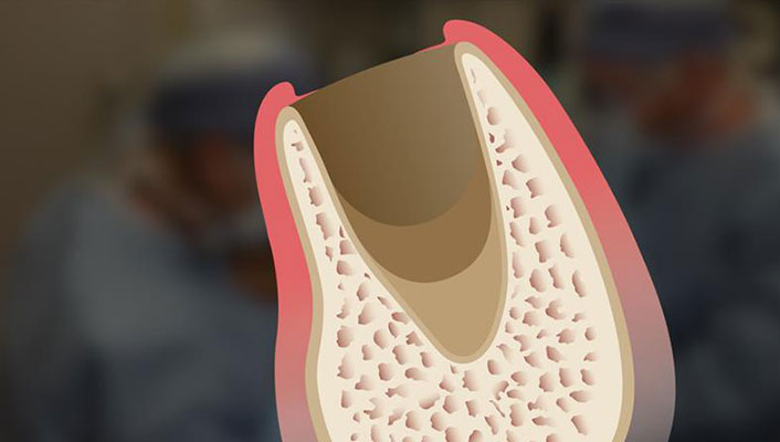
- Classifying the extraction site based on the number of walls remaining
- How to determine if bone grafting is indicated
- Treatment options determined by extraction socket walls
- Predictable treatment of extraction sockets
- The five stages of extraction socket healing
- Contraindications to socket grafting
Extraction and Socket Grafting: Part 2—Extraction Site Healing
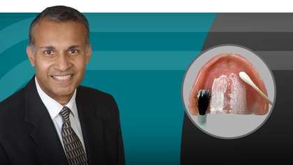
Presenter
Course Objectives
1 CE Credit
Dentists have seen significant advancements in the understanding of bone healing at the cellular and molecular level, which has led to techniques that have expanded the scope of oral implantology. In most cases, the extraction process will initiate a sequence of bony resorptive morphologic changes that may negatively alter the alveolar ridge. These changes are variable and, if significant, can eliminate the possibility of future implant placement. Therefore, it is crucial to understand the processes of repair and regeneration, and to apply their concepts to diagnosis and treatment planning with respect to the treatment of extraction sites. In this article, Dr. Resnik discusses extraction site healing, along with indications and contraindications for socket grafting.
Topics include:
Summary
A classification of the indications for socket grafting has been discussed with regard to the socket walls remaining after tooth extraction. When bony walls are thick in a five-walled extraction socket, grafting is optional. However, when all walls of the socket are present but one or more walls are thin, socket grafting is indicated. When additional walls are missing, socket grafting is highly recommended. In general, the more walls compromised or missing, the greater the need for grafting. When clinicians thoroughly understand how bone healing occurs, treatment of the extraction site can be more predictable and successful for patients.
Recognition & Approval
Glidewell Education Center
Nationally Approved PACE Provider for FAGD/MAGD credit
Approval does not imply acceptance by any regulatory authority, or AGD endorsement. 3/1/2024 to 2/29/2028.
Provider ID# 216789
References
- Misch CE, Suzuki JB. Tooth extraction, socket grafting, and barrier membrane bone regeneration. In: Misch CE, editor. Contemporary Implant Dentistry, 3rd ed. St. Louis: Mosby; 2008. p. 870-904.
- Ohta Y. Comparative changes in microvasculature and bone during healing of implant and extraction sites. J Oral Implantol. 1993;19(3):184-98.
- Vera C, De Kok IJ, Reinhold D, Limpiphipatanakorn P, Yap AK, Tyndall D, Cooper LF. Evaluation of buccal alveolar bone dimension of maxillary anterior and premolar teeth: a cone beam computed tomography investigation. Int J Oral Maxillofac Implants. 2012 Nov-Dec;27(6):1514-9.
- Artzi Z, Tal H, Dayan D. Porous bovine bone mineral in healing of human extraction sockets. Part 1: histomorphometric evaluations at 9 months. J Periodontol. 2000 Jun;71(16):1015-23.
- Wang HL, Kiyonobu K, Neiva RF. Socket augmentation: rationale and technique. Implant Dent. 2001 Dec;13(4):286-96.
- Fishel D, Buchner A, Hershkowith A, Kaffe I. Roentgenologic study of the mental foramen. Oral Surg Oral Med Oral Pathol. 1976 May;41(5):682-6.
- Kang SH, Kim BS, Kim Y. Proximity of posterior teeth to the maxillary sinus and buccal bone thickness: a biometric assessment using cone-beam computed tomography. J Endod. 2015 Nov; 41(11):1839-46.
-
 Online CE CourseBone Grafting: Selecting Biomaterials Based on the Anatomical DefectUnderstand the best practices for choosing the appropriate bone grafting material based on the anatomical defect that is present.
 -
 Online CE CourseComplete Dentures Part 4: Room for ImprovementUnderstanding the indications and maintenance requirements of implant overdentures is key to improving retention. Dr. Raymond Choi explains the characteristics of optimal retention, from setting proper expectations for patients to identifying recommended procedures to increase success.
 -
 Online CE CourseComplete Dentures Part 3: Delivery DayMaximize delivery success by integrating proven techniques for comfortable and esthetic dentures. Dr. Sree Koka demonstrates and discusses essential steps such as try-ins, patient instruction, overdenture attachment pick-up techniques and occlusal adjustments.